X-rays
X-radiation (composed of X-rays) is a form of electromagnetic radiation. Most X-rays have a wavelength ranging from 0.01 to 10 nanometres, corresponding to frequencies in the range 30 petahertz to 30 exahertz and energies in the range 100 eV to 100keV.
X-ray wavelengths are shorter than those of UV rays and typically longer than those of gamma rays. In many languages, x-radiation is referred to with terms meaning Röntgen radiation, after Wilhelm Röntgen, who is usually credited as its discoverer, and who had named it X-radiation to signify an unknown type of radiation.
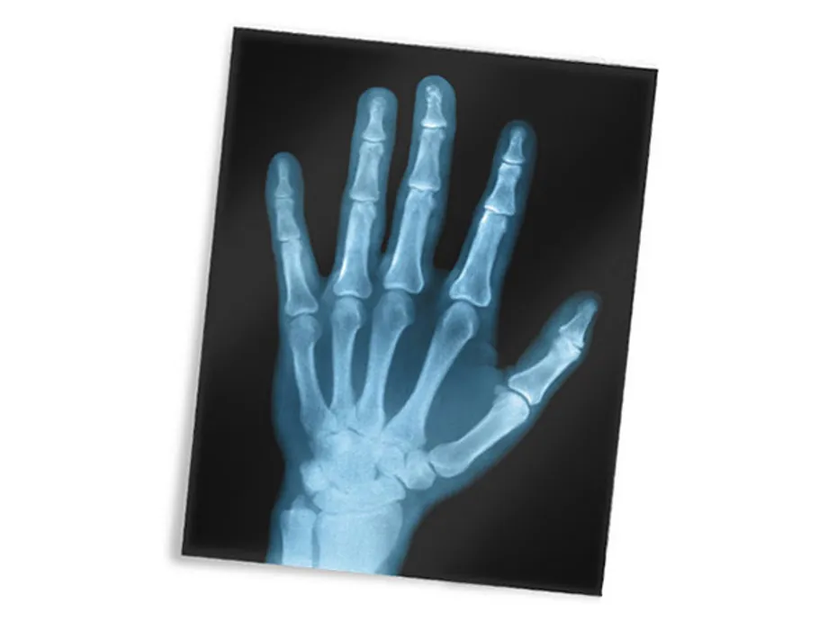
X-ray image of hand
This image, or radiograph, is a modern X-ray of a hand. One of the first X-ray photographs was made of the hand of Röntgen's wife.
The image displayed both her wedding ring and bones. A radiograph is an X-ray image obtained by placing a part of the patient in front of an X-ray detect or and then illuminating it with a short X-ray pulse.
Bones contain much calcium, which due to its relatively high atomic number absorbs X-rays efficiently. This reduces the amount of X-rays reaching the detector in the shadow of the bones, making them clearly visible on the radiograph.